

Dr Mannan Gupta
Medically Reviewed by Dr. Mannan Gupta On June 19, 2026
You saw blood. Your heart dropped. And now you’re sitting with an ultrasound report that says “subchorionic hematoma” a term nobody explained to you properly.
If you’re searching for subchorionic hematoma treatment in New Delhi and feeling overwhelmed, you’re not alone and you’ve come to the right place.
This condition is more common than most people realize, and the majority of women who receive this diagnosis go on to have healthy pregnancies. But you deserve a clear, honest explanation — not vague reassurances.
Key Takeaways
- What a subchorionic hematoma actually is and why it happens
- How serious it can be with real numbers, not fear
- What treatment options exist and what the evidence says
- How to manage daily life safely during this diagnosis
- What your chances of a healthy pregnancy actually look like
What Exactly Is a Subchorionic Hematoma?
Medical Definition in Plain Language
A subchorionic hematoma is a collection of blood between the wall of the uterus and the chorion the outer membrane that surrounds your developing baby.
Think of it as a small pocket of blood that has pooled in a space where it shouldn’t be. It is also called a subchorionic hemorrhage.
Where Does the Blood Collect?
The chorion is the membrane that eventually forms part of the placenta. When it partially separates from the uterine wall, blood fills that gap.
This separation is what shows up on an ultrasound as a dark, crescent-shaped area near the gestational sac.
How Common Is It Really?
Subchorionic hematoma is the most common cause of bleeding in the first trimester, occurring in approximately 1–3% of all pregnancies. In women who experience first-trimester bleeding, the rate is significantly higher studies from PubMed put it closer to 18–20%. It is not rare. It is not a sign that something catastrophic has happened.
For women in New Delhi experiencing first-trimester bleeding and seeking expert evaluation, Pregnancy Care in New Delhi at Dr. Mannan IVF Centre includes early pregnancy ultrasound and specialist review exactly the kind of prompt assessment this diagnosis requires.
Does Size Matter?
Yes and this is something many doctors do not explain clearly enough. Small hematomas (under 25% of the gestational sac volume) carry a much lower risk than large ones.
Large hematomas, particularly those that form near the placenta rather than away from it, require closer monitoring.
What Causes a Subchorionic Hematoma?
The Implantation Theory
The most widely accepted explanation is that during implantation when the embryo attaches to the uterine wall a small area of the membrane lifts slightly, causing local bleeding. This is considered a mechanical event, not a failure of your body.
The IVF and Assisted Reproduction Link
Women who conceive through IVF have a measurably higher incidence of subchorionic hematoma. Research published in reproductive medicine journals suggests this may be related to the hormonal environment created during stimulation, or to the embryo transfer process itself. If you’re an IVF patient, this is important context your doctor should have shared with you upfront.
Understanding the full picture of what a post-IVF pregnancy can involve is equally valuable our guide on High-Risk Pregnancy After IVF: Precautions covers the monitoring steps and warning signs that are specifically relevant for women who have conceived through assisted reproduction.
Hormonal and Clotting Factors
Low progesterone levels, uterine abnormalities, and underlying clotting disorders have all been associated with subchorionic hemorrhage in pregnancy. Women on blood thinners or with conditions like antiphospholipid syndrome may face higher risk.
What Doesn’t Cause It?
Exercise, sex, stress, or lifting something heavy did not cause your hematoma. This is not your fault. This is a structural event that happens during early development, and no amount of rest beforehand would have reliably prevented it.
Dr Mannan IVF Centre
Experience world-class fertility care with Dr. Mannan Gupta at the Best IVF Centre in Delhi
How Will You Know If You Have One?
Symptoms That Bring Women to the ER
Vaginal bleeding in the first trimester is the symptom that sends most women to emergency rooms or their obstetrician immediately. The blood may be bright red, brown, or pink. Mild cramping may accompany it. These symptoms are alarming and they deserve immediate evaluation but they do not automatically mean pregnancy loss.
If you are trying to understand whether your bleeding means your pregnancy is classified as high-risk, our article on whether bleeding in pregnancy is always a sign of a high-risk pregnancy provides a clear, evidence-based answer including when bleeding is reassuring and when it genuinely warrants escalation.
Silent Hematomas No Symptoms at All
A significant number of subchorionic hematomas are found incidentally — meaning a routine ultrasound picks them up with zero symptoms. The woman had no bleeding, no pain. This is actually more common than most people realize, and it underscores why regular ultrasound monitoring in early pregnancy matters.
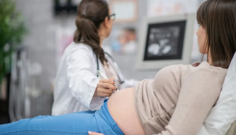
How Ultrasound Confirms the Diagnosis?
A transvaginal or abdominal ultrasound is the only reliable way to diagnose this condition. The hematoma appears as a hypoechoic (darker) crescent area near the gestational sac. Your doctor should measure the hematoma and document its location relative to the placenta — both factors affect prognosis significantly.
How Serious Can It Get — And When Should You Worry?
Risk Factors That Increase Severity
Large hematoma size, a hematoma located beneath the placenta (subplacental), advanced maternal age, and underlying clotting disorders all increase the risk of complications.
Being informed about your specific risk profile is your right as a patient — ask your doctor directly.
Link to Miscarriage — Real Numbers, Not Fear
Studies show that small subchorionic hematomas resolve on their own in over 80% of cases with no adverse outcome. Even with larger hematomas, many pregnancies continue successfully.
The presence of a fetal heartbeat alongside a hematoma is a strongly reassuring sign. However, large subplacental hematomas do carry a statistically elevated risk of first-trimester pregnancy loss, and your doctor should be honest with you about that.
Second Trimester Risks
If a hematoma persists into the second trimester, risks include placental abruption, preterm labor, and premature rupture of membranes. This is not inevitable — but it does require closer surveillance with more frequent ultrasounds.
When It’s Likely to Resolve Safely
Most hematomas diagnosed in the first trimester, particularly small ones away from the placenta, resolve by 20 weeks. The body reabsorbs the blood gradually. Serial ultrasounds are the only reliable way to confirm this is happening.
What Treatment Options Are Actually Available?
Why There Is No Universal Drug Protocol?
This is the part your doctor may have glossed over: there is currently no drug that has been proven in large randomized controlled trials to definitively treat subchorionic hematoma. This does not mean nothing can be done it means management is individualized and evidence-based clinical judgment matters enormously.
Progesterone Supplementation Evidence and Debate
Many doctors prescribe vaginal or oral progesterone to women with subchorionic hematomas, particularly those who conceived via IVF. The rationale is that progesterone supports uterine lining stability and may reduce further separation.
While definitive large-scale trial data is still emerging, many reproductive specialists consider it a reasonable low-risk intervention, especially in progesterone-deficient patients.
Bed Rest What the Research Actually Says
Strict bed rest has not been shown in controlled studies to directly resolve hematomas faster. However, reducing strenuous physical activity, avoiding intercourse, and limiting travel are commonly recommended as precautionary measures to avoid additional mechanical stress on an already vulnerable area. Watchful waiting with activity modification is the standard of care in most cases.
What Doctors Monitor and How Often?
Monitoring typically involves serial ultrasounds every 1–2 weeks to assess hematoma size, fetal heart rate, and placental position. If the hematoma is growing, or if fetal heart rate becomes abnormal, the management plan must be escalated immediately.
For women in New Delhi navigating this diagnosis, having a care team experienced in high-risk pregnancy monitoring is essential you can learn more about the structured surveillance approach available through High-Risk Pregnancy Care in New Delhi and what a specialist-led plan for this condition looks like in practice.
If you are navigating this diagnosis and want expert guidance from a specialist who works with high-risk and IVF pregnancies daily, Dr. Mannan Gupta at Dr. Mannan IVF Centre has supported many women through exactly this situation. You can explore care options and book a consultation at drmannanivfcentre.com.
What Does Daily Life Look Like With This Diagnosis?
Activity Restrictions – What to Avoid
Avoid high-impact exercise, heavy lifting, and prolonged standing. Walking gently is generally acceptable unless your doctor has specifically advised full bed rest. Listen to your body increased bleeding after activity is a clear signal to stop and call your doctor.
Sexual Activity and Travel
Sexual intercourse should be avoided until the hematoma has resolved on ultrasound. Long-haul air travel carries dehydration and thrombosis risks that are best avoided during this period. Discuss any planned travel with your treating doctor before booking anything.
Warning Signs to Act On Immediately
Go to the emergency department immediately if you experience heavy vaginal bleeding (soaking a pad in an hour), severe abdominal pain, fever, or dizziness. These are not symptoms to monitor at home.
Emotional Toll and Mental Health
This diagnosis creates weeks sometimes months of uncertainty. Anxiety, grief, and fear are completely normal responses. If you find yourself unable to function or experiencing symptoms of prenatal depression, speak to your doctor. Psychological support during high-risk pregnancy is not a luxury it is part of your care
Can You Have a Normal Pregnancy After This?
Outcomes Data — Most Cases Resolve
The evidence is genuinely encouraging. The majority of women diagnosed with a subchorionic hematoma, particularly those with small-to-medium hematomas and a confirmed fetal heartbeat, deliver healthy babies. A 2011 meta-analysis in Obstetrics & Gynecology found that while subchorionic hematoma increases risk of certain complications, most pregnancies proceed without major incident.
Follow-Up Care and Fetal Monitoring
After resolution, your pregnancy should continue with standard or slightly enhanced monitoring depending on your history. Confirm with your doctor whether additional anomaly scans or Doppler flow studies are recommended given your specific case.
Planning Future Pregnancies
A history of subchorionic hematoma does not automatically mean it will recur. However, if you have an underlying clotting disorder or conceived via IVF, a pre-pregnancy evaluation with a reproductive specialist is strongly advisable before your next cycle.
If you have also experienced a previous pregnancy loss, it is worth knowing that subchorionic hematoma is one of several uterine and vascular factors that can contribute to recurrent loss — our article on repeated miscarriage and the hidden causes most doctors miss outlines the full investigation pathway that should follow more than one loss, including the clotting and uterine assessments most directly relevant to your situation.
Final Thoughts
A subchorionic hematoma diagnosis is frightening but it is not a death sentence for your pregnancy. Most hematomas resolve. Most pregnancies with this diagnosis result in healthy babies.
What matters most is accurate diagnosis, honest communication from your care team, appropriate monitoring, and sensible activity modification.
You deserve a doctor who explains your situation clearly, tracks your progress closely, and adjusts your care plan based on evidence not assumptions. Don’t settle for vague reassurances or dismissive consultations.
Frequently Asked Questions
1. Can I still have a healthy baby if I have a subchorionic hematoma?
Yes and this is the most important thing to understand. The majority of women diagnosed with this condition, especially those with small hematomas and a confirmed fetal heartbeat, go on to deliver healthy babies. Your doctor should be giving you this information clearly, not just monitoring you silently.
2.How long does a subchorionic hematoma take to heal?
Most small-to-medium hematomas resolve within 4–8 weeks. Larger hematomas may take longer or persist into the second trimester. Serial ultrasounds are the only reliable way to track resolution do not assume it has healed without imaging confirmation.
3. Should I be on complete bed rest with a subchorionic hematoma?
Full bed rest has not been proven to speed up resolution, but reduced activity is widely recommended. Most doctors advise avoiding strenuous exercise, intercourse, and heavy lifting. Ask your specific doctor what level of restriction is appropriate for your hematoma size and location.
4. Is there a difference between a subchorionic hematoma and placental abruption?
Yes these are distinct conditions. A subchorionic hematoma is blood between the uterine wall and the outer fetal membrane, typically diagnosed early in pregnancy. Placental abruption is when the placenta itself separates from the uterine wall, which is a more serious emergency typically occurring later in pregnancy. They can sometimes be confused on reports, so ask your doctor to clarify exactly what was seen.
5. Does a subchorionic hematoma mean I will miscarry?
No it increases statistical risk slightly, but it does not predict miscarriage. Many women with this diagnosis including those with larger hematomas carry their pregnancies to full term. The key factors are hematoma size, location, fetal heart rate, and how closely your pregnancy is monitored going forward.
